
Mohs Micrographic Surgery
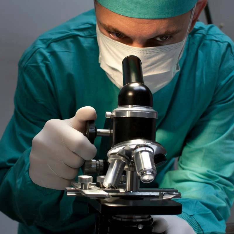
We are proud to offer cutting-edge Mohs micrographic surgery at our Port Arthur and Victoria, Texas locations, delivering exceptional skin cancer treatment with precision and care. Our on-site CLIA-certified high-complexity laboratories ensure seamless access to:
Expertise
Our skilled Mohs surgeons deliver precise and compassionate care.
Convenience
Timely and accurate diagnoses are made possible with on-site labs, eliminating the need for external facility visits.
Comprehensive care
Our advanced labs enable meticulous tissue examination and cancer cell detection, guaranteeing optimal treatment outcomes.
By combining Mohs surgery with on-site CLIA-certified labs, Gemini Dermatology offers unparalleled care and peace of mind, all within the comfort of our local communities.
What is Mohs Micrographic Surgery?
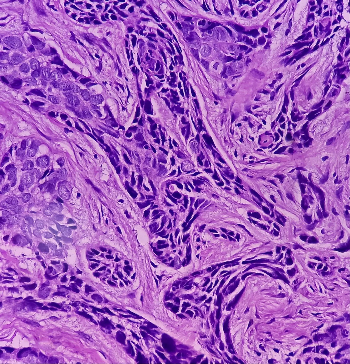
Mohs micrographic surgery is a highly effective and precise tissue sparing surgical technique used to remove skin cancers including basal cell carcinomas, squamous cell carcinoma, and other rare skin cancers.
Developed by Dr. Frederic Mohs in the 1930s, Mohs surgery was initially used to treat skin cancer with a zinc chloride fixative. Through continuous innovation and improvement, Mohs micrographic surgery has emerged as a cornerstone of skin cancer treatment, trusted for its precision and outstanding results.
Could Mohs Surgery be for me?
- Skin cancers in sensitive areas, such as the head, neck, face, nose, lips, eyelids, hands, feet, or genitals, where scarring could have a significant cosmetic or functional impact.
- Recurrent skin cancers that have returned after previous treatments.
- Large skin cancers (greater than 2.0 cm) located anywhere on the body.
- Skin cancers in patients with weakened immune systems, such as those with autoimmune disorders or organ transplant recipients.
- Cases where preserving healthy tissue and minimizing scarring is crucial.
How does Mohs Surgery work?
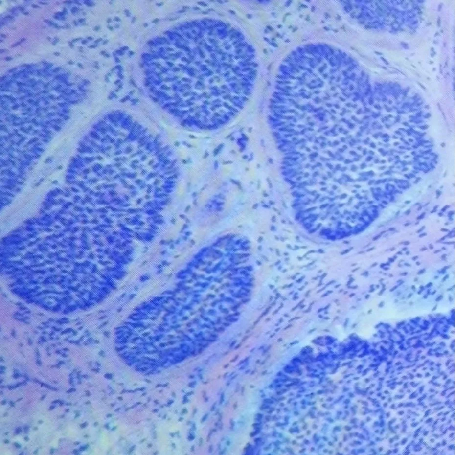
Mohs surgery involves removing the visible tumor and a thin layer of surrounding tissue. The removed tissue is then taken to an on-site laboratory, where it is processed and converted into a slide using a cryostat. This specialized equipment rapidly freezes and sections the tissue, allowing for immediate examination. The slide is then handed to the Mohs surgeon, who interprets it under a microscope to determine if cancer cells are still present. If cancer cells are found, the process is repeated, with the Mohs surgeon removing additional tissue and examining it under the microscope until all cancerous tissue is removed.
Once the cancer is clear, the Mohs surgeon uses reconstructive techniques, incorporating advanced facial plastic surgery skills, to close the defect in a way that minimizes scarring and allows for the best cosmetic and functional outcomes. These techniques, which are an integral part of Mohs training, may involve suturing, grafting, flaps, or other specialized methods to restore the natural appearance and function of the affected area. By combining meticulous cancer removal with expert reconstruction, Mohs surgery offers a comprehensive approach to skin cancer treatment, ensuring optimal results for patients all with the benefit of minimal to no scarring.
Mohs Micrographic Surgery FAQ
-
- The procedure takes place in the convenient outpatient setting of our clinic exam room.
- A relatively painless local anesthetic is administered to ensure your comfort during the procedure.
- Following each stage, unwind in our waiting room, featuring amenities designed to ease your wait and make the experience as stress-free as possible
- Once you're in the clear, we'll invite you back into the room to explore personalized closure options that align with your unique needs and desired results.
- Leave at the end of the day, confident in your skin cancer-free status.
-
In most cases, yes. (In some cases the closure will be performed at an outside facility by a head and neck surgeon).
Mohs surgery training typically includes reconstruction plastics training in the following areas:
1. Wound closure techniques: Sutures, staples, and adhesive closure
2. Skin grafting: Full-thickness and split-thickness grafts
3. Local flaps: Advancement, rotation, and transposition flaps
4. Facial reconstruction: Eyelid, nose, lip, and ear reconstruction
5. Scar revision: Techniques for optimizing scar appearance
6. Tissue rearrangement: Z-plasty, M-plasty, and other tissue rearrangement techniques
7. Facial aesthetics: Understanding facial anatomy and aesthetics to optimize reconstruction outcomes
Mohs surgeons receive training in these areas to enable them to perform reconstruction procedures immediately following Mohs surgery, ensuring optimal cosmetic and functional outcomes for their patients.
Combining Mohs surgery with post-Mohs reconstructive repairs offers a powerful approach to skin cancer treatment, delivering exceptional results: natural-looking outcomes, preserved function, and minimized scarring. With a remarkable cure rate approaching 99%, this integrated approach ensures the best possible outcomes for patients, providing both effective treatment and optimal cosmetic and functional results.
-
- High cure rate (up to 99%)
- In-office, same-day procedure for convenience and efficiency
- Minimally invasive, preserving healthy tissue
- Minimal scarring
- Unlike most surgeries, no PRE-OP fasting
- Highly effective for aggressive or recurrent skin cancers
- Restores natural appearance and function
- Optimal outcomes, even in sensitive or large areas
- Comfortable atmosphere with TV, snacks, and refreshments to reduce stress and promote relaxation
-
- Reduces reliance on radiation, decreasing the risk of side effects such as:
• Radiation burns
• Scarring
• Increased risk of secondary cancers
• Cosmetic disfigurement
- Accessible, specialized care for skin cancer patients
- Reduced travel time and costs for patients
- Enhanced patient satisfaction
- Supports comprehensive skin cancer management
- Fosters collaboration between dermatologists and other healthcare professionals